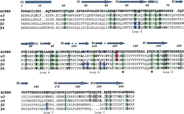
Figure 1.
Sequence alignment between AChBP and amino-terminal domains of rat α3, α4, β2 and β4 nAChR subunits. In this figure, the numbering of amino-acid residues refers to AChBP only. The top line (blue) presents the secondary structure of AChBP: bars indicate α-helices and arrows β-sheets. Green, gray, blue and red colors indicate residues present in all subunits, residues forming the agonist-binding site, those forming the antagonist-binding site, and residue interacting with the allosteric ligand, respectively. Asterisks indicate the beginning and the end of the Cys loop. The loops indicated in the bottom line are referred to residues belonging to the agonist-binding site.

