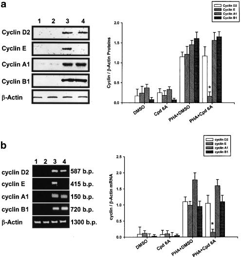
Figure 4.
Inhibition of cyclin E production and gene expression in T lymphocytes by Cpd 6A. T cells (1 × 10⁷) were stimulated with PHA (5 μg ml−1) in the presence or absence of 25 μM Cpd 6A for 24 h. Lysates (50 μg of protein) were run on a 10% SDS–PAGE gel and analyzed by immunoblotting with anti-Cyclin D2, E, A1 or B1 antibody (a). The total cellular RNA was isolated from T cells (5 × 10⁶) treated with Cpd 6A for 18 h and the RT–PCR was done as described in Methods. Following the reaction, the amplified product was taken out of the tubes and run on 2% agarose gel (b). Unstimulated cells (lane 1), unstimulated cells treated with Cpd 6A (lane 2), stimulated cells (lane 3), and stimulated cells in the presence of Cpd 6A (lane 4). The results are representative of three experiments. Bar graphs represent the ratio of each cyclin to β-actin signal. *P<0.001 vs the cells treated with PHA and DMSO.

