Figure 1.
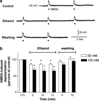
Concentration-dependent inhibition by ethanol of NMDA-induced depolarizations in SPNs of spinal cord slices. (a) a Representative continuous recording shows a sustained inhibition of NMDA-induced depolarization by ethanol (100 mM) superfused for 15 min in a sympathetic preganglionic neuron. The neuron was held at a resting membrane potential of −55 mV. The membrane depolarizations were induced by consecutive superfusions of NMDA (50 μM, filled circles) for 7 s at intervals of 5 min in the presence of TTX (0.5 μM). Downward deflections are hyperpolarizing electrotonic potentials induced by constant hyperpolarizing current pulses (not shown). (b) Graph shows percentage change in NMDA-induced depolarization against time in minutes following superfusion of two concentrations of ethanol (50, 100 mM) for 15 min and after removal of ethanol. The magnitude of NMDA-induced depolarization immediately prior to application of ethanol is taken as control (CTL) of 100%. The NMDA-induced depolarizations were 18.2±1.3 mV (n=7) and 17.6±0.9 mV (n=6) before the superfusion of 50 and 100 mM ethanol, respectively. *Significant difference from control.

