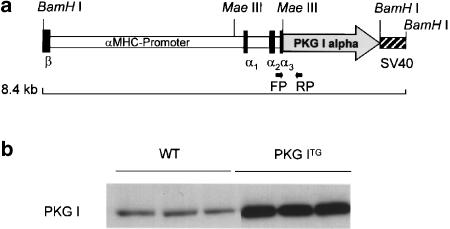
Figure 1.
Generation of transgenic mice overexpressing PKG I alpha in cardiac myocytes. (a) Human PKG I cDNA was placed under the control of murine αMHC promoter. PKG ITG mice were identified by genomic PCR amplification of a 513 bp fragment. Forward (FP) and reverse primers (RP) are indicated. (b) Protein expression of PKG I (76 kDa) was determined in cardiac ventricles of WT and in PKG ITG mice by Western-blot analysis using specific antisera and a peroxidase-labelled goat anti-rabbit antibody in an ECL detection system. To obtain immunoreactive signals in the linear range, 20 μg protein from WT hearts and 2 μg protein from PKG ITG hearts were loaded. PKG I expression was significantly upregulated in PKG ITG compared to WT ventricles by ∼46-fold (mean±s.e.m., n=13 per genotype, *P<0.01 vs WT).

