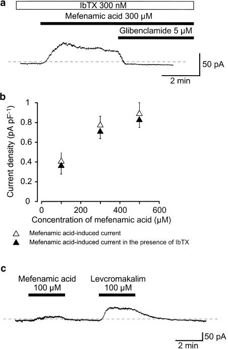
Figure 2.
Effects of mefenamic acid on membrane currents in pig urethral myocytes. The bath was superfused by PSS and the pipette solution was 140 mM KCl, containing 5 mM EGTA in a conventional whole-cell recording. (a) Under voltage-clamp conditions, in the presence of bath-applied 300 nM IbTX, 300 μM mefenamic acid caused an outward current at −50 mV. Glibenclamide (5 μM) completely suppressed this current. The dashed line indicates the zero current level. (b) Relationships between the peak amplitude of the mefenamic acid-induced outward currents and the concentration of mefenamic acid in the absence or presence of 300 nM IbTX (the bath) at −50 mV. Each symbol indicates the mean of 5–8 observations with s.d. shown by vertical lines. (c) The comparative effects of 100 μM mefenamic acid and l00 μM levcromakalim on membrane current. The dashed line indicates the zero current level. Extracellular application of 100 μM mefenamic acid caused an outward membrane current at −50 mV. About 2.5 min later, levcromakalim caused a larger amplitude outward current with a similar basal noise level.

