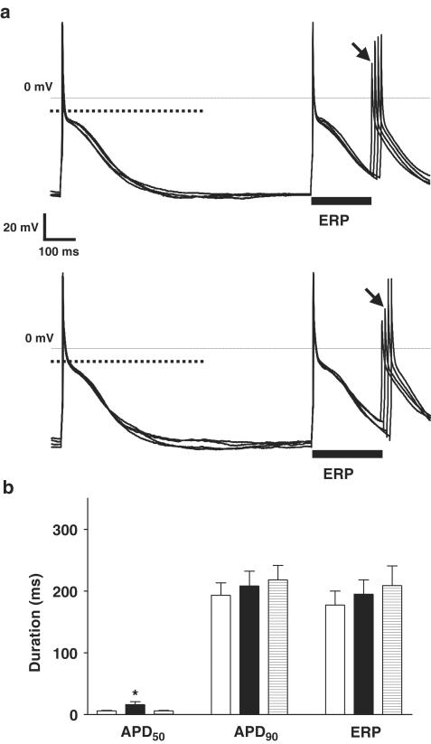
Figure 4.
Effect of 5-HT on action potentials and refractoriness in single human atrial cells from patients not treated with β-blockers. (a) Representative examples of original action potential recordings obtained from a single human atrial myocyte, from a patient not treated with a β-blocker, before (upper panel) and in the presence of 10 μM 5-HT (lower panel). Cells were paced at 75 bpm. Dotted lines in bold show the level of 50% of the action potential amplitude. The majority of cells displayed type 3 action potentials, that is, with pronounced phase 1 and a plateau amplitude below the 50% level. The ERP, indicated by solid bars, was calculated as the longest S1–S2 interval failing to elicit an S2 response of amplitude >80% of the preceding S1 action potential. The S2 response used to measure this interval is labelled with an arrow. (b) Mean (±s.e.m.) APD (ms) measured at 50 and 90% repolarisation (APD50 and APD90, respectively; n=16 cells, eight patients) and ERP (n=12 cells, eight patients) in cells from patients not treated with β-blockers, in the absence (open bars), in the presence (closed bars) and after removal of 10 μM 5-HT (striped bars; n=11 cells, eight patients for APD and n=9 cells, seven patients for ERP). Asterisk denotes P<0.05 between control and 5-HT values (paired t-test).

