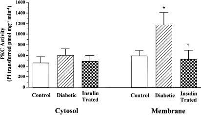
Figure 8.
PKC activities in partially purified cytosolic and membranous fractions from control, diabetic, and insulin-treated diabetic rat hearts. PKC activity was measured as the rate of transfer of 32P from [γ-32P]ATP into the specific substrate in the presence of Ca2+, phosphatidylserine, and phorbol 12-myristate 13-acetate. Values are means±s.e.m. of six separate experiments. *Significantly different from control (P<0.05). †Significantly different from diabetic (P<0.05).

