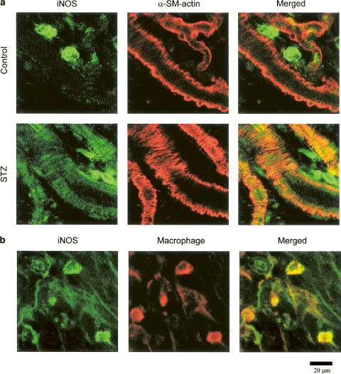
Figure 4.
Immunohistochemical localization of iNOS in the mesenteries of control and STZ-diabetic rats. (a) The mesenteries isolated from the control (upper panels) and the STZ-diabetic rats (lower panels) were double-immunolabeled for iNOS (green fluorescence; left panels) and α-smooth muscle (SM) actin (red fluorescence; center panels). (b) The mesenteries isolated from the control rats were double-immunolabeled for iNOS (green fluorescence; left panel) and macrophage (red fluorescence; center panel). The yellowish area in each right panel is produced by the coincidence of both fluorescences.

