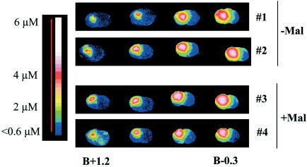
Figure 3.
I.str. distribution of [3H]M826. Rats 1 and 2 received an i.str. infusion of M826 in the absence of prior injection of malonate (Mal−). Rats 3 and 4 received an infusion of M826 10 min after injection of malonate (Mal+) in the same striatum. Both malonate and M826 were injected in the left striatum as described in Methods. All four animals were euthanized 1 h after infusion of M826. Coronal cryostat sections of the brains were processed for autoradiography. The sections were color coded from blue (weakest concentration) to white (strongest concentration).

