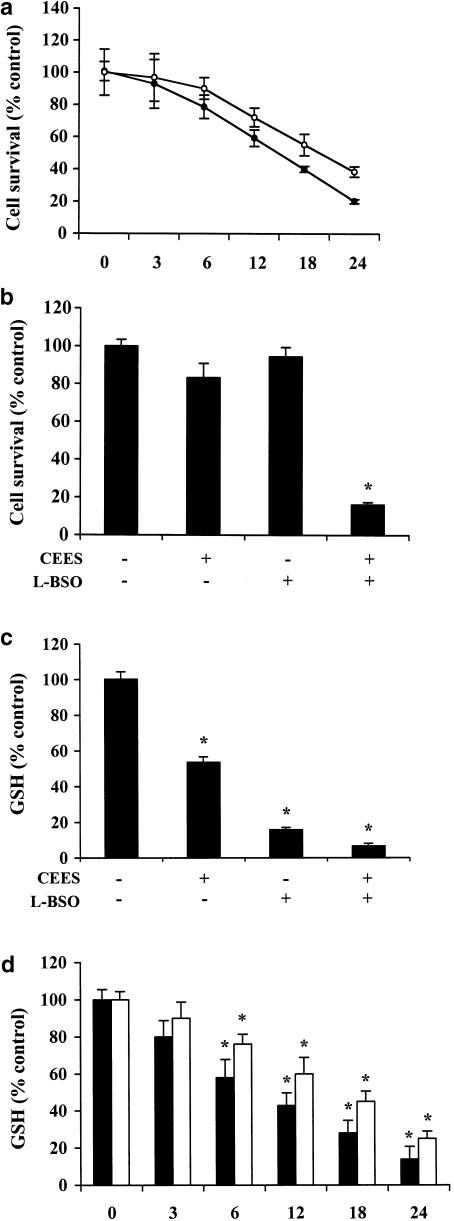
Figure 1.
Role of intracellular GSH in CEES-induced death in lymphocytes. (a) Jurkat cells (closed circles) and normal lymphocytes (open circles) were incubated for the indicated times with 600 μM CEES, after which cell viability was assessed by measurement of calcein-AM fluorescence. Jurkat cells were incubated first for 20 h with or without 200 μM L-BSO and then for 6 h in the additional absence or presence of CEES, after which cell viability (b) and intracellular abundance of GSH (c) was determined. (d) Jurkat cells (closed bars) and lymphocytes (open bars) were treated with 600 μM CEES at the indicated times, after which GSH content was analysed. All data are means±s.d. of triplicates from an experiment that was repeated a total of three times with similar results. (*)P<0.05 versus control value for untreated cells.

