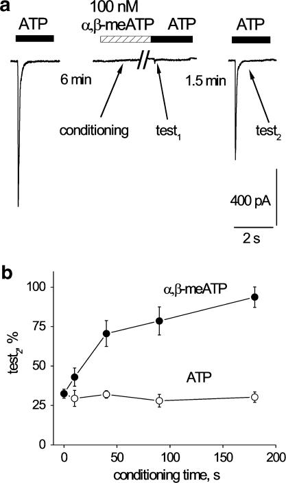
Figure 6.
Duration of application of a small concentration of agonist shapes recovery of response to ATP of DRG neurons. (a) Example of protocol to test low concentration of agonist (100 nM α,β-meATP) on ATP responses. Conditioning agent, which does not induce any measurable membrane current, is switched off immediately before applying ATP and yet it fully desensitizes the subsequent test1 response. Test2 response is generated at a fixed 1.5 min interval after test1 to probe recovery from desensitization. (b) Length of conditioning application of 100 nM ATP (open circles) or 100 nM α,β-meATP (filled circles) versus amplitude of test2 responses evoked by 10 μM ATP. Note that the amplitude of test2 responses is uniformly depressed by ATP conditioning (n=5–7 cells). Conversely, conditioning with 100 nM α,β-meATP largely facilitates recovery of test2 responses, a phenomenon dependent on the length of α,β-meATP conditioning (n=4–6 cells).

