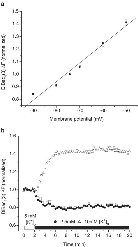
Figure 1.
Effects of diastolic membrane potential (Em) and selected [K+]o on DiBac4(3) fluorescence in right ventricular myocytes isolated from adult rat hearts. (a) Cardiomyocytes were voltage-clamped using the perforated patch-clamp technique. At selected membrane potentials, changes in DiBac4(3) fluorescence intensity (ΔF) were assessed and ΔF was used as an estimate of relative Em changes in subsequent imaging experiments. ΔF was normalized in each cell to F measured at the resting membrane potential, which averaged –74.25±0.86 mV (n=5 cells). The line was fitted by linear regression. (b) Note that 2.5 mM [K+]o hyperpolarized, and 10 mM [K+]o depolarized the Em of quiescent, normoxic myocytes.

