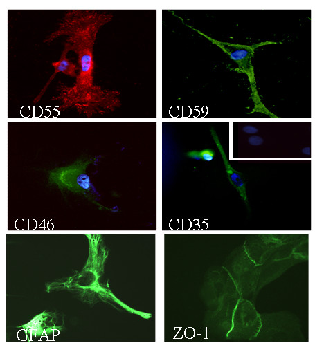
Figure 2.

Immunofluorescence analyses of the human tumour-derived ependymoma primary cultures (clone 9945) stained for complement regulatory proteins and cell markers. Cells on coverslips were fixed with acetone and stained with antibodies against ependymal cells specific markers (GFAP, ZO-1 and S100, not shown) and complement regulators proteins (CD55, CD59, CD46 and CD35). Original magnification ×400. Background staining was observed using irrelevant antibodies (inset). Nuclei were counterstained with DAPI (blue).
