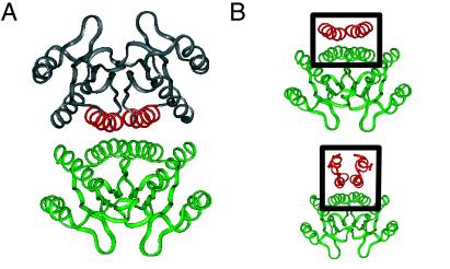
Figure 4.
Structure of transcriptional coactivator DCoH (PDB ID code 1dch; refs. 22–24) (A) and model of the DCoH-HNF-1α complex (B). (A) The DCoH homotetramer is formed by an antiparallel dimer of saddles (upper and lower dimers). The lower dimer is shown in green relative to binding helices (red) of upper dimer (gray). (B Upper) The tetramer interface of DCoH contains an antiparallel 4HB (box) with dihedral (D2) symmetry. (Lower) Side view of proposed model of HNF-1α (residues 5–31; red in box) atop the DCoH dimer (green). The predicted interface's symmetry differs from that of the DCoH-DCoH tetramer.

