
Figure 1.
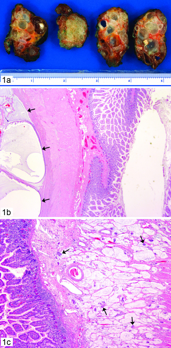
Case 1: 1a. Pseudomyxoma peritonei with multilobulated polypoid gelatinous masses, 1b. Subserosal cystic structures filled with mucin and lined by single layer of bland epithelium (arrow) in the small bowel. (hematoxylin-eosin, × 10); 1c. Numerous cell clusters floating in the mucin pools and infiltrating the submucosa of small bowel (arrow) (haematoxylin-eosin, × 20)- PMCA variant of PMP