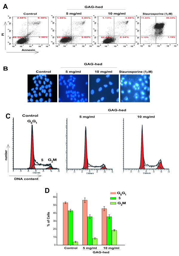
Figure 5.

GAG-hed arrest cell cycle but did not induce apoptosis. (A) 1 × 10⁶ cells were treated with GAG-hed (5 and 10 mg/ml) for 48 h. The cells were then permeablized, stained for Annexin V, and stored on ice until analyzed by FACS. The upper panels show representative examples of control HeLa cells either untreated (left) or treated with staurosporine to induce apoptosis (right). Increased Annexin V staining was seen in HeLa cells only in the presence of staurosporine. In all panels, cells in the lower left quadrant are alive, cells in the lower right quadrant are in early apoptosis, in the upper right are in late apoptosis, and cells in the upper left quadrant are dead. Percentage of total signal within the quadrant is indicated. (B) Fluorescent microscopic analysis of cells stained with DAPI. Forty-eight hours after GAG-hed treatment the cells were fixed, stained with DAPI and analyzed for morphological characteristics associated with apoptosis. (C) Cell cycle distribution in HeLa cells treated with GAG-hed. Histograms are derived from a single experiment that was repeated three times with similar results, that in (D) are expressed as percentages for each of the three cell cycle phases, mean ± SE of three independent experiments.
