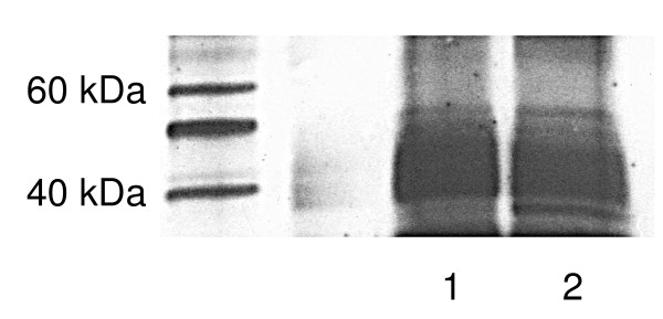
Figure 2.
Immuno-precipitation to determine that the lower MW protein in Figure 1 was GIRK1. Two samples of MDA-MB-453 cells were immuno-precipitated with goat polyclonal antibody (Santa Cruz) for GIRK1, then separated by Western blot, and probed with rabbit polyclonal antibody used for Figure 1 (Upstate). The highest expression of GIRK1 was at the lower molecular weight in these immuno-precipitated cells. This is a representative gel of two separate experiments.

