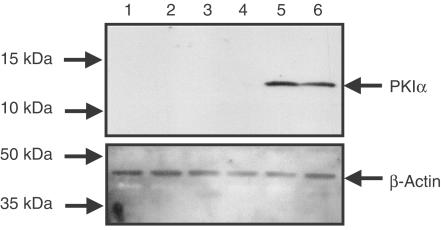
Figure 5.
Expression of PKIα in HASM cells infected with Ad5.CMV.PKIα. Adherent cells were cultured until 50% confluent and then infected with Ad5.CMV.Null, Ad5.CMV.PKIα (MOI=100) or left untreated (naïve) for 48 h at 37°C. Cells were growth arrested in serum-free medium and processed by western blotting for PKIα expression (a) and the house-keeping protein, β-actin (b). Data are representative of three independent determinations using tissue from different donors. See Methods for further details. (Key: lanes 1 & 2, Naïve; lanes 3 & 4, Ad5.CMV.Null; lanes 5 & 6, Ad5.CMV.PKIα).

