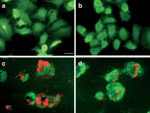
Figure 2.
Fluorescent images of calcein-AM (green) and propidium iodide (red) double-stained cells. Tumor cells were grown on poly-L-lysine or Collagen IV-coated coverslips, exposed to Taxol for 48 h, and subsequently analyzed for apoptosis as described in Methods. (a) A549 control, (b) A549-T24 control, (c) A549 treated with 20 nM Taxol, (d) A549-T24 treated with 200 nM Taxol. Scale bar=25 μm.

