Figure 3.
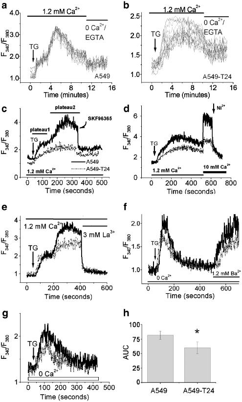
Steady-state and agonist-induced intracellular calcium levels in tumor cells. The cells were loaded with Fura-2 AM and [Ca2+]c was detected as described in Methods. (a, b) Single-cell calcium measurements by fluorescence imaging microscopy in TG (100 nM)-treated A549 (a) and A549-T24 cells (b), followed by removal of extracellular calcium. [Ca2+]c changes were recorded in 15–20 cells. (c–e) A representative tracing of the response of [Ca2+]i of A549 and A549-T24 cells in response to TG (100 nM), followed by SKF96365 (50 μM) treatment (c), addition of extracellular calcium (10 mM) and Ni2+ (10 mM) treatment (d), or La3+ (3 mM) treatment (e). (f) A representative tracing of the response of [Ca2+]i of A549 and A549-T24 cells in response to TG (100 nM) in Ca2+-free medium, followed by addition of 1.2 mM Ba2+. (g) A representative tracing of the response of [Ca2+]i of A549 and A549-T24 cells in response to TG (100 nM) in the absence of extracellular calcium. (h) The mean±s.d. of AUC of TG (100 nM) response in the absence of extracellular Ca2+. *Significantly different (P<0.01, N=6) from A549 by Mann–Whitney test.

