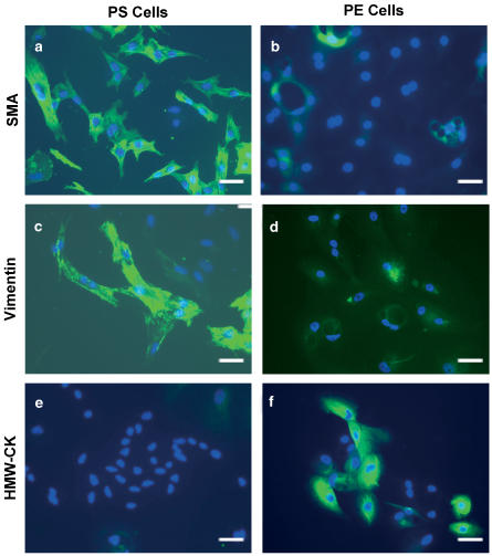
Figure 1.
Primary cultures of canine PS and PE cells. PS (a, c, e) and PE (b, d, f) cells were labeled with SMA, vimentin or HMW-CK. Nuclear staining is indicated by the blue DAPI labeling. Data shown were taken at × 20 magnification and is representative of three independent experiments. The horizontal bar represents 5 μm.

