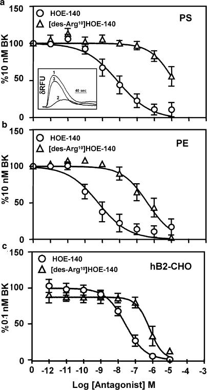
Figure 3.
Antagonism of BK-induced calcium mobilization in canine PS, canine PE and hB2-CHO cells. Representative FLIPR traces, from PS cells (similar for PE and hB2-CHO cells), displaying the kinetics of BK (10 nM)-induced intracellular Ca2+ mobilization measured as changes in δRFU (trace 1) in the presence of 10 nM HOE-140 (trace 2) or 10 nM [des-Arg10]HOE-140 (trace 3) are shown as an inset to Figure 3a. Increasing concentrations of HOE-140 and [des-Arg10]HOE-140 were used to antagonize BK-induced FLIPR responses in canine PS (a), canine PE (b) and hB2-CHO (c) cells. Responses were normalized to 10 nM BK (a and b) or 0.1 nM BK (c). Points represent mean values and vertical lines indicate standard errors of the mean from four to six independent experiments.

