Figure 2.
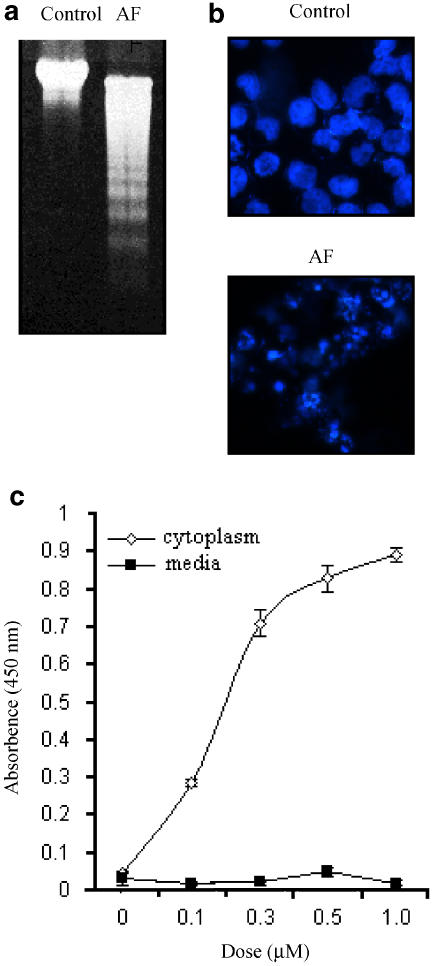
AF-induced DNA fragmentation in NB4 cells. (a) The NB4 cells were incubated with medium alone (control) or 1 μM AF for 24 h. Genomic DNAs were isolated and analysed on a 1% agarose gel. (b) The AF-treated cells were stained with Hoechst 33342 (10 μM) for 30 min, and then observed under a fluorescence microscope equipped with a DAPI filter. (c) BrdU-labelled NB4 cells (1 × 10⁴ cells) were incubated for 6 h in various concentrations of AF (0–1 μM). The BrdU-labelled DNA fragments in the cytoplasm or culture media were quantitated using cellular DNA fragmentation ELISA kit (Roche, Germany). Experiments were carried out in triplicate and the results represent means±s.d.

