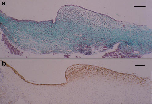
Figure 2.
Fetal lamb at 0.7 gestation. Longitudinal section of the ductus venosus showing the inlet region with a sphincter-like formation and a segment of the vessel. (a) Masson's trichrome staining. (b) α-Actin immunostaining. The muscle component was less developed than in the term preparation but, otherwise, it was similarly arranged. Bar 100 μm.

