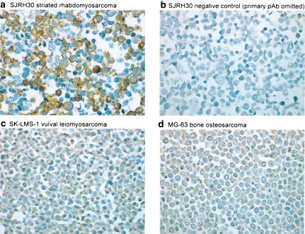
Figure 4.
hUT immunoreactivity is detectable in SJRH30 cells using a novel affinity-purified rabbit anti-hUT pAb. (a) Globular/granular staining is observed at the plasma membrane of formalin-fixed, paraffin-embedded SJRH30 cell pellets labeled with 1.0 μg ml−1 hUT pAb. (b) In contrast, staining of the SJRH30 cell pellet is abolished following preincubation of the SJRH30 cells with anti-hUT pAb in the presence of antigen (1 h at 5 μg ml−1). Similarly, no membrane staining is noted in the ‘non-striated' human sarcoma cell lines prepared in an identical manner, including (c) SK-LMS-1 (vulval leiomyosarcoma) and (d) MG-63 (bone osteosarcoma) cell lines.

