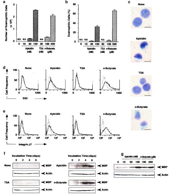
Figure 1.
Differentiation of HL-60 clone 15 cells into eosinophils induced by apicidin, TSA and n-butyrate. HL-60 clone 15 cells were incubated in the presence of apicidin, TSA or n-butyrate. (a, b) The number (a) and the percentage (b) of eosinophilic cells after 6 days incubation in the presence of the indicated concentrations of apicidin, TSA or n-butyrate. N.D., not detectable. (c) Microscopic observation of HL-60 clone 15 cells incubated for 6 days in the presence of apicidin (100 nM), TSA (30 nM) or n-butyrate (500 μM). The bar indicates 10 μm. (d, e) The intracellular structure (d) and the expression of integrin β7 (e) after 6 days incubation in the presence of apicidin (100 nM), TSA (30 nM) or n-butyrate (500 μM). Dotted and solid lines represent the histogram before and after incubation with the drug, respectively. (f, g) The expression of MBP in the cells incubated for the periods indicated in the presence of apicidin (100 nM), TSA (30 nM) or n-butyrate (500 μM) (f) or for 6 days in the presence of the indicated concentrations of apicidin or n-butyrate (g). Actin in the cell lysate was detected by immunoblotting as an internal control.

