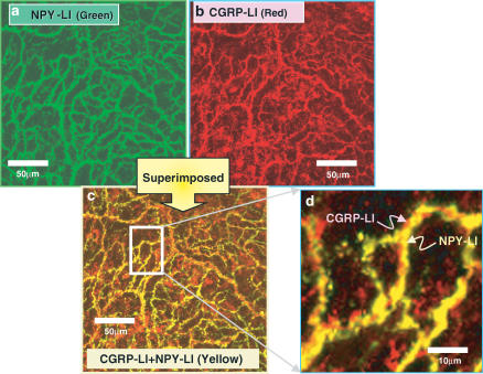
Figure 5.
Confocal laser photomicrograph showing NPY (a; green)-like immunoreactive (LI)- and CGRP (b; red)-LI-containing fibers in the mesenteric artery. (a) and (b) are photographs of the same area in the whole mount of the artery. (c) is a superimposed image of (a) and (b). Colocalization of CGRP-LI and NPY-1-LI is recognized as yellow in many fibers. (d) shows higher magnification of the rectangular area in (c). The scale bar in (c) and (d) indicates 50 and 10 μm, respectively.

