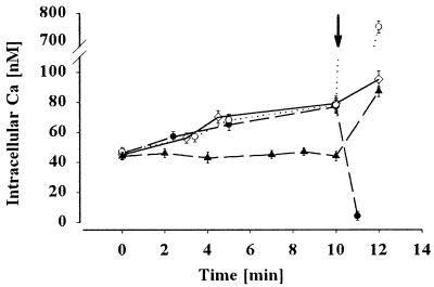
Figure 6.
Effect of the Ca2+ mobilizer thapsigargin on the intracellular Ca2+concentration of the quiescent cells. Sperm were loaded with calcium green1-AM as described in Materials and Methods and suspended in NoCaFPS (closed circle) or FPS containing 100 nM Ca2+ (open diamond) or 1,000 nM Ca2+ (open circle), and 50 μM thapsigargin and fluorescence from the cells was monitored for 10 min. Control cells were suspended in FPS containing 100 nM Ca2+ (closed circle), and at t = 10 min, 5 μM A 23187 calcium ionophore was added to the suspension and the fluorescence from the sperm cells was measured. When 5 μM A 23187 was added to the sperm cells containing different calcium concentrations and thapsigargin (arrow), the intracellular calcium concentration reached that of the extracellular value.

