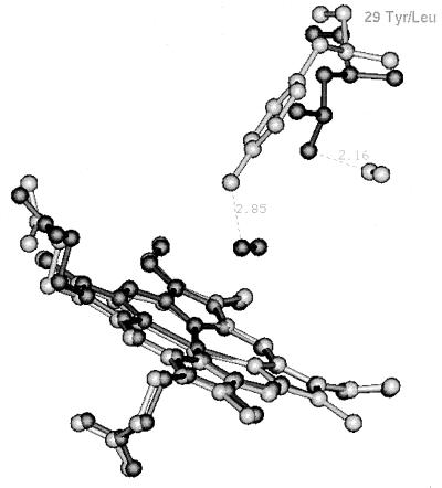
Figure 3.
The position of CO* in wt Mb and Mb-YQR, relative to residue at B10. Superposition of wt Mb⋯CO* (8) (dark) and [Mb-YQR⋯CO*] (light). For both proteins the model shows the heme, the photolyzed CO*, and the residue at position B10 i.e., Leu for wt Mb and Tyr for Mb-YQR. We also show: (i) the distance (2.9 Å) expected between the hydroxyl group of Tyr(B10)29 and the photolyzed CO* if this was to occupy the primary docking site (dark), characteristically observed in wt Mb; and (ii) the distance (2.2 Å) expected between Leu(B10)29 and the photolyzed CO* in the secondary docking site (light) identified in the photolyzed intermediate of Mb-YQR (see Fig. 1B).

