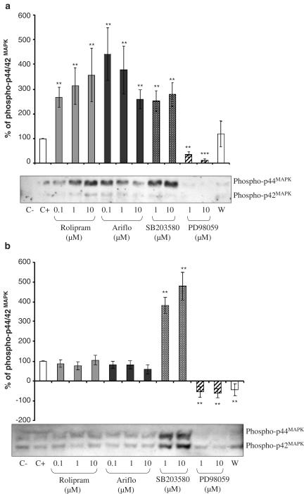
Figure 5.
(a) Western blot of phosphorylated p44/42MAPK and percentage of phosphorylated p44/42MAPK evaluated by densitometry (OD mm−2) of the blots in BAL neutrophils of LPS-treated rats 10 min after 2 μM fMLP addition in the presence of PDE4 inhibitors or MAPK inhibitors (C−=negative control, C+=positive control, W=0.05 nM wortmannin). (b) Western blot of phosphorylated p44/42MAPK and percentage of phosphorylated p44/42MAPK evaluated by densitometry (OD mm−2) of the blots in BAL macrophages of LPS-treated rats at the activation peak induced by 2 μM fMLP in the presence of PDE4 inhibitors or MAPK inhibitors (C−=negative control, C+=positive control, W=0.05 nM wortmannin).

