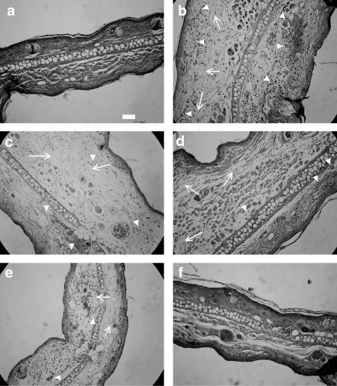
Figure 4.
Histological appearance of mouse ear (left ear), 8 h after the induction of dermatitis (panels b–f) or of naïve animals (panel a). Mice received a topical treatment (5 min before or after the induction of dermatitis) with vehicle (panel b), pre- and post-hydrocortisone (panels c and d, respectively) or pre- and post-NCX 1022 (panels e and f, respectively). Hydrocortisone and NCX 1022 were used at the dose of 3 nmol. Scale bar: 50 μm.

