
Fig. 6.
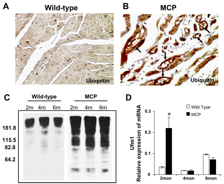
Accumulation of ubiquitinylated proteins in the hearts of wild-type and MCP mice. Immunohistochemical staining of ubiquitin in the hearts of wild-type controls (A) and MCP (B) mice of 6 mo of age. Note the absence or weaker cytoplasmic staining (brown) in cardiomyocytes of wild-type controls and the strong staining in cardiomyocytes undergoing degenerative changes such as vacuolation (arrow). Original magnification, ×400. C: Western blotting of heart homogenates from wild-type controls and MCP mice of 2, 4, and 6 mo of age showing marked accumulation of ubiquitinylated proteins in the hearts of MCP mice. D: elevated expression of ubiquitin fold modifier 1 (Ufm1) in the hearts of MCP mice determined by quantitative real-time PCR. *Significantly different from wild-type controls (P < 0.05).