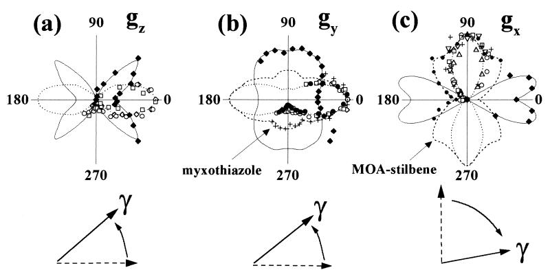
Figure 2.
Orientations of the paramagnetic axes of the Rieske cluster with respect to the membrane plane as measured by the dependences of signal size on the angle between the magnetic field and the membrane. The three signals denoted gx,y,z (see also Fig. 1) correspond to the principal directions of the paramagnetic center (19). Samples oriented in the absence (open diamonds, dotted lines) and in the presence of the inhibitors stigmatellin (open squares, dotted lines), UHDBT (open circles, dotted lines), myxothiazole (crosses, dashed lines) and MOA–stilbene (full circles, dashed lines) were studied. The control sample was furthermore examined with oxidized (open up-triangles, dotted lines) or reduced (open down-triangles, dotted lines) quinone in the Qo-site (Fig. 2c), giving rise to gx-troughs at differing field positions. Signal amplitudes measured on the irradiation-induced center are denoted by continuous lines and filled diamonds (the gx-signal was evaluated on the UHDBT-treated sample; see text). (Lower) Schematic representation of the directions of these three axes in the chemically (dotted lines) and the irradiation-reduced (continuous lines) states.

