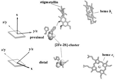
Figure 6.
Schematic representation of the Rieske cluster's magnetic axes with respect to the membrane in the chemically (Upper) and the irradiation-reduced (Lower) state as compared with the two different (proximal and distal) conformational states observed in the crystal structures (Protein Databank entries 3bcc and 1bcc). Only heme bL, heme c1, the inhibitor stigmatellin occupying the Qo-site, and the two conformational states of the [2Fe-2S]-cluster of the ISP (together with the two ligating histidine residues) are shown.

