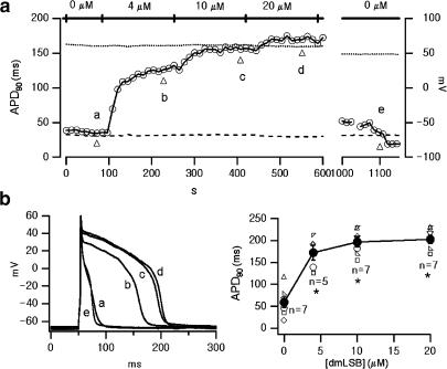
Figure 2.
Effects of dmLSB on cardiac AP. (a) Change in AP duration during the bath application of dmLSB. All data points were obtained from the same cell. Horizontal bars above the plot indicate the duration of the bath application of dmLSB. APs were evoked by depolarizing current pulse (55 pA, 5 ms, 2 Hz). APD90 (AP duration at 90% repolarization) was obtained from the average of five sequential APs selected every 12 s. (b) Left, exemplary AP records at various concentration of dmLSB (a: control; b: 4 μM; c: 10 μM; d: 20 μM; e: wash out of dmLSB). The time point of each AP record was indicated by a triangle in (a). Right, APD90 as a function of the concentration of dmLSB. APD values obtained from seven different cells were superimposed (smaller open symbols). At each concentration of dmLSB, a mean value for APD90 was also superimposed (larger closed circles; error bars, s.e.m.). Asterisks indicate statistical significance (paired t-test; P<0.01).

