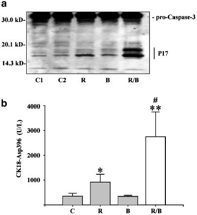
Figure 2.
Activation of caspase-3 by ritonavir plus butyrate in DLD-1 cells. (a) DLD-1 cells were kept as unstimulated control (C1), as vehicle control (C2, 0.1% DMSO), or were stimulated with ritonavir (R, 60 μM), butyrate (B, 5 mM), or ritonavir plus butyrate (R/B). After 20 h of stimulation, DLD-1 cells were harvested and homogenates were assayed for caspase-3 protein by immunoblot analysis. One representative blot of two independently performed experiments is shown. Total protein (100 μg) was applied per lane. (b) After 20 h of stimulation, cell-free culture supernatants were assayed for the presence of the cytokeratin-18 neoantigen CK18-Asp396 by ELISA. Data are shown as mean CK18-Asp396 concentrations±s.d. (n=4). Vehicle control (0.1% DMSO). *P<0.05 compared to unstimulated control; **P<0.01 compared to unstimulated control; #P<0.05 compared to ritonavir alone.

