Figure 4.
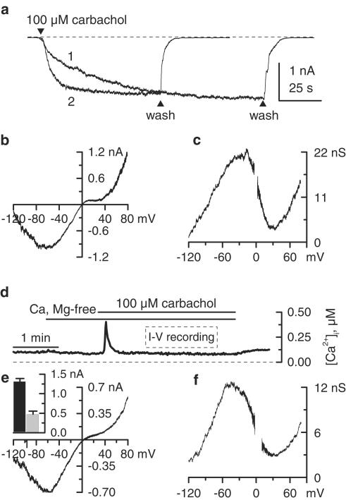
Possible mICAT inhibition by a diffusible intracellular factor. (a) Delayed current activation when carbachol was applied immediately after break-through (trace 1; note that PSS has been already replaced by high-Cs+ external solution before establishing the whole-cell configuration) compared to standard 3 min equilibration experiment (trace 2). (b, c) Lack of intracellular ATP (20 mM) effect on the 100 μM carbachol-induced mICAT I–V relationship and corresponding cation conductance activation curve. Cell dialysis with PA-free pipette solution containing 1 mM ATP appears to be sufficient to remove PAs from the cytoplasm. (d) Changes of [Ca2+]i in single ileal smooth muscle cell voltage-clamped at −40 mV during the application of 100 μM carbachol in Ca2+-free high-Cs+ external solution. I–V relationships in perforated patch experiments were measured when [Ca2+]i was stabilized at about 100 nM, as indicated. (e, f) Carbachol (100 μM) induced mICAT I–V relationship and corresponding cation conductance activation curve measured using perforated patch configuration. Mean current amplitudes using this (grey column, n=7) and conventional whole-cell configuration (black column, n=12) are shown in the inset.

