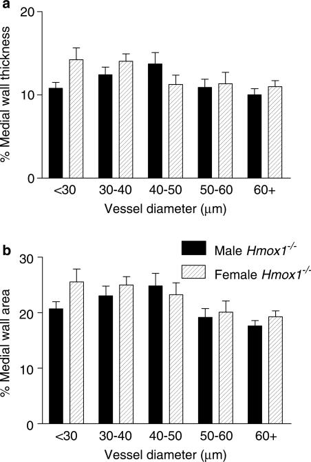
Figure 10.
Pulmonary vascular structure in adult male versus adult female, HO-1 deficient mice. Lung tissue harvested from adult male Hmox1−/− mice and adult female Hmox1−/− mice was fixed in 10% formol saline, embedded in paraffin wax, sectioned (4 μm) and stained using an antibody specific for smooth muscle α-actin (dilution 1 : 3000) using the streptavidin–biotin–peroxidase complex method. Images were captured using a Zeiss AxioCam and analysed using Openlab. The external and internal diameter, and area, of muscular and partially muscular pulmonary arteries was measured. Percentage medial wall thickness/area was then calculated. (a) Percentage medial wall thickness and (b) percentage medial wall area of arteries from adult male Hmox1−/− (22–41 vessels from n=6 animals were analysed), and adult female Hmox1−/− (12–38 vessels from n=5 animals were analysed), mice. Data are mean±s.e.m. No significant difference (two-way ANOVA) was observed between the two groups.

