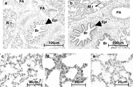
Figure 2.
Localisation of HO-1 in the porcine lung. Porcine lung tissue was fixed in 10% formal saline, embedded in paraffin wax, sectioned (4 μm) and stained with an antibody specific for HO-1 (dilution 1 : 500) using the streptavidin–biotin–peroxidase complex method. Figure showing HO-1 expression in (a) foetal and (b) 14-day-old porcine lung. In photographs of the same lung sections, at a higher magnification, panels (c) and (d) show the alveolar region of the foetal and 14-day-old animals, respectively. Panel (e) shows the negative control (no primary antibody) using tissue from the same 14-day-old animal. PA, pulmonary artery; Br, bronchus; Epi, epithelium; Al r, alveolar region. Slides are representative of sections studied from the three other animals in each age group.

