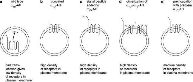
Figure 3.
Schematic illustration of α1D-adrenoceptor surface expression in transfected cells. The wt α1D-AR is poorly translocated to the plasma membrane (a). Truncated α1D-AR variant (b) and the α1D-AR supplemented with a cleavable signal peptide (c) are well expressed in the membranes. Coexpression of α1D-AR with the α1B-subtype increases surface expression of α1D-adrenoceptors (d). The well-expressed receptors (b–d) show a six to 10-fold increase in the density of binding sites in the plasma membrane compared to the wt α1D-AR. Incubation of α1D-AR-expressing cells with the α1-antagonist prazosin induces an increased density of receptors in the plasma membrane (e). The figure is based on results reported in the present study (c), and in Pupo et al. (2003) (a and b), Hague et al. (2004b) (d), and McCune et al. (2000) (e).

