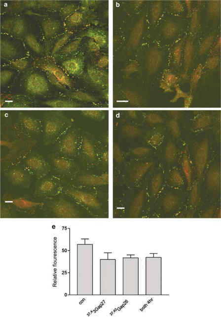
Figure 4.
Integrity of gap junction plaques in A7r5 cells fixed and stained for Cx43 (green) and Cx40 (red) following 4 h incubation with connexin-mimetic peptides. (a) Control, (b) 37,43Gap 27 (600 μM), (c) 37,40Gap 26 (600 μM), (d) 37,43Gap 27+37,40Gap 26 (300 μM each). (e) Histogram showing plaque integrity quantified by analysis of Cx43 and Cx40 fluorescence at the plasma membrane subtracted from background fluorescence following the various treatments. Results are given as mean relative fluorescence±s.e.m. Bars=10 μm. Magnification × 40.

