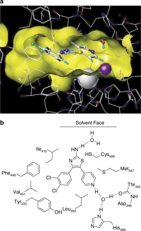
Figure 3.
(a) Model of CGH2466 docked in the PDE4 active site looking from the solvent into the binding pocket. The ligand is drawn in a ball and stick representation, protein and water molecules in a wire frame representation and the metal ions as spheres. The surface of the binding site is shown in yellow. Parts of the surface have been removed for clarity. (b) Schematic representation of the binding mode of CGH2466 in PDE4.

