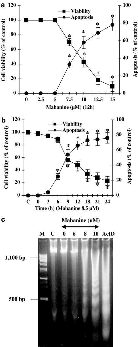
Figure 2.
Mahanine inhibits cell proliferation and induces apoptosis in U937 cells. (a and b) Dose and time dependent response of U937 cells to mahanine. Cells were incubated with the indicated concentration of mahanine for 0–24 h. The data show that mahanine increased number of apoptotic cells and decreased cell viability in both experiments. Cell viability was determined using the trypan-blue exclusion assay, and the number of apoptotic cells was counted after staining with Hoechst 33258. Data are mean values from three separate experiments and bars indicate standard deviations. *P<0.05 as compared with controls. (c) Mahanine-induced DNA fragmentation in U937 cells. Cells were incubated with the indicated amount of mahanine for 10 h. DNA was extracted as described in ‘Methods' and visualized on agarose gels. M, marker DNA; C, cells incubated with media alone; ActD, cells incubated with actinomycin D (positive control).

