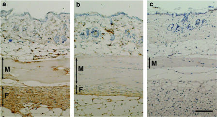
Figure 4.
Effect of a chymase inhibitor on immunohistochemical staining of TGF-beta1. Histological cross-sections of the skin tissues from vehicle-treated Tsk mouse (a), SUN-C8257-treated Tsk mouse (b), and vehicle-treated control C57BL/6J mouse (c) were incubated with antibody against TGF-beta1, and then the tissue-bound antibody was detected with the DAKO ENVISION kit and DAB substrate. Sections were then counterstained with hematoxylin. The brown color staining indicates the presence of TGF-beta1. F: fibrous layer; M: panniculus carnosus muscle layer. Scale bar=100 μm.

