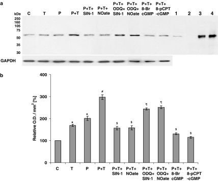
Figure 5.
Western analysis of NADPH oxidase in PAECs using a monoclonal antibody directed against the gp91phox subunit of mouse macrophage NADPH oxidase. Cells were either not treated or stimulated overnight with either prednisolone (P; 1 μM) or TNF-α (T; 10 ng ml−1) alone or with combination of the two (±SIN-1 (100 nM); DETA-NONOate (NOate; 500 μM); SIN-1+ODQ (1 μM); DETA-NONOate+ODQ; 8-bromo-cGMP (100 μM) and 8-pCPT-cGMP (100 μM)). Where applied, PAECs were preincubated with ODQ for at least an hour before SIN-1 or DETA-NONOate was added. Cellular lysates of stimulated mouse macrophages (lane 1), pig neutrophils (lane 3) and human neutrophils (lane 4) were used as positive controls. Whole-cell lysate of PAVSMCs (lane 2) was used as an internal control and GAPDH expression as a loading control. Panel a shows the representative blot and panel b the results of the densitometric analyses of six blots (expressed as relative optical density (OD) mm−2). *P<0.05, significantly increased compared to control. #P<0.05, significantly increased compared to prednisolone/TNF-α-treated cells only. $P<0.05, significantly inhibited compared to prednisolone and TNF-α-treated cells. ¶P<0.05, significantly enhanced compared to cells treated with SIN-1 or DETA-NONOate alone in the presence of prednisolone and TNF-α.

