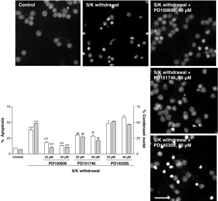
Figure 2.
Antiapoptotic effect of PD150606, PD151746 and PD145305 on S/K withdrawal-induced changes in the percentage of cells rated as apoptotic by means of flow cytometric (hypodiploid cells, open bars) or morphological analysis (gray bars). Results are show as mean±s.e.m. of 4–6 independent cultures. Statistical analysis was carried out by one-way ANOVA followed by Tukey's test; ***P<0.001 vs control values; ###P<0.001 vs S/K withdrawal values. Representative fluorescence photomicrographs showing chromatin condensation in permeabilized CGNs in the different experimental conditions. The nuclei were counted at the fluorescence microscope, distinguishing the normal from the condensed nuclei with the criteria stated in Methods. Calibration bar, 10 μm.

