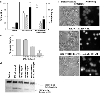
Figure 3.
Analysis of calpain and caspase-3 activities in 12 h S/K withdrawal in CGNs in the absence or presence of PD150606 (40 μM), PD151746 (40 μM) and z.VAD.fmk (100 μM). (a) Effect of z.VAD.fmk (100 μM) on S/K withdrawal-induced changes in the cell viability (open bars) or in the percentage of cells rated as apoptotic (hypodiploid cells/dashed bars; condensed nuclei/black bars). (b) Representative phase contrasts and fluorescence photomicrographs of CGNs in the different experimental conditions. Calibration bar, 10 μm. (c) Caspase-3 activity in CGNs after 12 h S/K withdrawal in the presence or absence of tested compounds. Results are the mean±s.e.m. of three cultures. The statistical analysis was carried out with the one-way ANOVA followed by Tukey's test, **P<0.05 vs S/K withdrawal values. (d) Representative Western blot of α-spectrin digestion showing calpain activity (SBPD145kD fragment) and caspase 3 activity (SBPD120kD fragment), influence of preincubation with 40 μM PD150606 or PD151746 and 100 μM z.VAD.fmk.

