Figure 1.
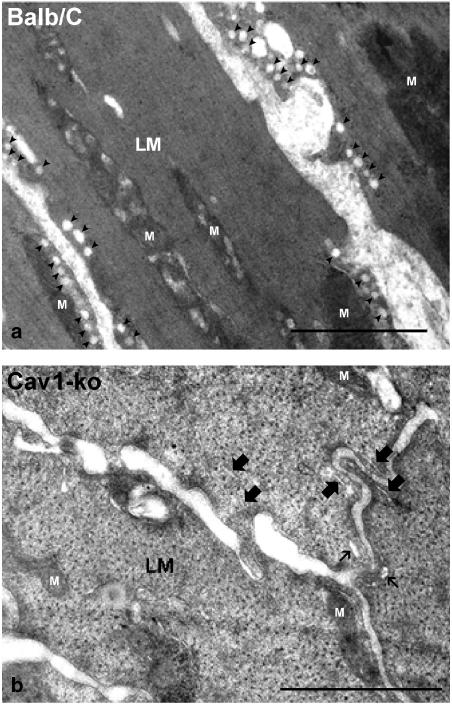
An electron microscope image of the longitudinal muscle layer (LM) of mouse small intestine. Note the total absence of the flask-shaped caveolae on the plasma membrane of the longitudinal muscle cells of Cav1−/− (b) as compared to the longitudinal muscle cells of the control BALB/c mouse (a). Caveolae are indicated by solid arrowheads. Small endoplasmic reticulum profiles (marked with thick arrows) and sarcoplasmic reticulum (marked with thin arrows) remain. Mitochondria are labeled with (M). Cav1+/+ intestine showed similar caveolae profile to the BALB/c (data not shown). Length bars are 1 μm.

