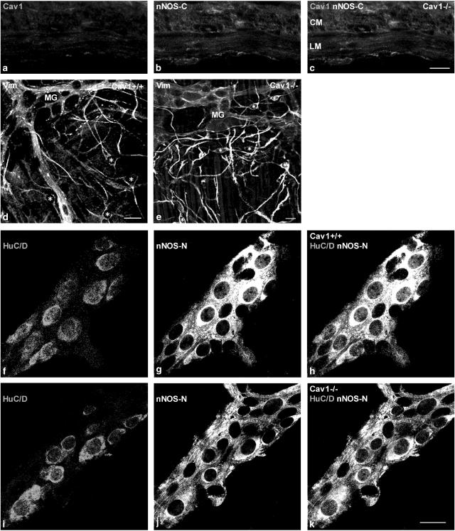
Figure 2.
Immunohistochemical staining of cryosections from Cav1−/− jejunum (a–c) and whole-mount preparations of myenteric plexus layer from Cav1+/+ and Cav1−/− jejunal tissues (d–k). Panels (a)–(c) show that there are no caveolin-1 and nNOS-C-immunoreactivities in all ICC and smooth muscles layers of Cav1−/− jejunum. Panels (d) and (e) show myenteric ICC stained with anti-vimentin in Cav1+/+ and Cav1−/− myenteric plexus, respectively. ICCs are marked with asterisks. Note that the myenteric plexus ICC are equally present and similarly distributed in both strains. Panels (f)–(k) show double staining of whole-mount preparations for myenteric neurons HuC/D protein and nNOS-N. Panels (f) and (i) show myenteric neurons stained with HuC/D in Cav1+/+ and Cav1−/− myenteric plexus, respectively. Panels (g) and (j) show myenteric neurons and nerve fibers stained with nNOS-N in Cav1+/+ and Cav1−/− myenteric plexus, respectively. Panels (h) and (k) show myenteric neurons co-localized with HuC/D and nNOS-N in Cav1+/+ and Cav1−/− myenteric plexus, respectively. Note the persistence of nNOS-N in myenteric neurons in Cav1−/− intestine. Length bars are 20 μm for panels (a)–(c), (d) and (e), and (f)–(k).

