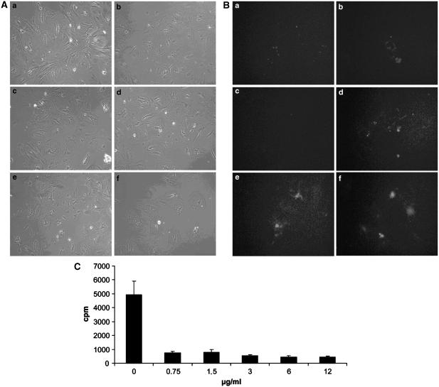
Figure 3.
Effect of increasing concentrations of cycloheximide on cultured HSC. Pictures a–f were taken 24 h after, respectively, 0, 0.75, 1.5, 3, 6 and 12 μg ml−1 of cycloheximide was added to the cultures. (A) At low concentration, cycloheximide had no visible effect on cell attachment (a–c). However, a concentration of 3 μg ml−1 cycloheximide or higher (d–f) resulted in detachment of cells. (B) Fluorescence microscopy of cells treated with propidium iodide after 24 h of treatment of cycloheximide confirmed these results. Definitely, at a concentration of 3 μg ml−1 or higher, cycloheximide induced apoptosis as demonstrated by fluorescent nuclei of apoptotic cells (d–f). (C) Determination of the dose of cycloheximide which effectively blocked protein synthesis. Cells were metabolically labelled with [35S]methionine/cysteine. Protein synthesis was assessed by trichloroacetic acid precipitation. From our data, it is evident that cycloheximide blocked protein synthesis by stellate cells, even at concentrations as low as 0.75 μg ml−1. Bars represent means±s.d. of three independent experiments.

